The Correctional Nurse should have a working knowledge of the primary lesion morphology (lesion types) that they may observe in the clinical area. Use of the proper terminology when describing the lesions seen is of upmost importance, especially when describing them to a Provider who is not onsite to observe themselves. Below find the most common lesion types.
Common Skin Lesions
Primary Lesions
First visible lesion or initial skin changes seen
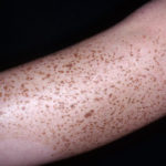
Macules are flat, nonpalpable lesions usually < 10 mm in diameter. Macules represent a change in color and are not raised or depressed compared to the skin surface.
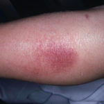
A patch is a large macule. Examples include freckles, flat moles, tattoos, and port-wine stains, and the rashes of rickettsial infections, rubella, measles, and some allergic drug eruptions.Patches may also have papules, non-palpable fine scale, and plaques.
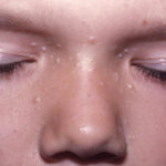
Papules are elevated lesions usually < 10 mm in diameter that can be felt or palpated. Examples include nevi, warts, lichen planus, insect bites, seborrheic keratoses, actinic keratoses, some lesions of acne, and skin cancers. Papule are typically further described by shape, size, color, and surface change.
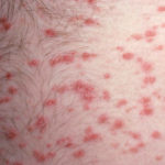
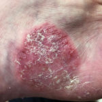
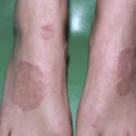
Plaques are palpable lesions > 10 mm in diameter that are elevated or depressed compared to the skin surface. Plaques may be flat topped or rounded. Lesions of psoriasis and granuloma annulare commonly form plaques.
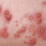
Vesicles are small, clear, fluid-filled blisters < 10 mm in diameter. Vesicles are characteristic of herpes infections, acute allergic contact dermatitis, and some autoimmune blistering disorders (eg, dermatitis herpetiformis).
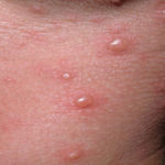
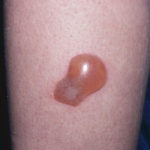
Bullae are clear fluid-filled blisters > 10 mm in diameter. These may be caused by burns, bites, irritant or allergic contact dermatitis, and drug reactions.
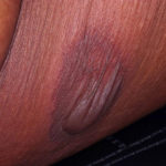
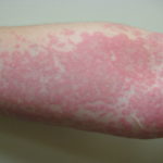
Urticaria (wheals or hives) is characterized by elevated lesions caused by localized edema. Wheals are pruritic and red. Wheals are a common manifestation of hypersensitivity to drugs, stings or bites. Autoimmune issues and physical stimuli, including temperature, pressure, and sunlight, may also cause urticaria. Typically the wheals last less than 24 hours.
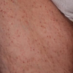
Petechiae are nonblanchable pinpoint areas of hemorrhage. Causes include platelet abnormalities (eg, thrombocytopenia, platelet dysfunction), vasculitis, and infections (eg, meningococcemia, Rocky Mountain Spotted Fever, other rickettsioses).
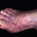
Purpura is a larger area of hemorrhage that may be palpable. Palpable purpura is considered the hallmark of leukocytoclastic vasculitis. Purpura may indicate a coagulopathy. Large areas of purpura may be called ecchymosis or bruise.
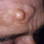
Telangiectases are areas of small, permanently dilated blood vessels that may occur in areas of sun damage, rosacea, systemic diseases (especially systemic sclerosis), or inherited diseases (eg, ataxia-telangiectasia, hereditary hemorrhagic telangiectasia) or after long-term therapy with topical fluorinated corticosteroids.